Wednesday 6 March 2013

Blogging from the cell sorter

10am
Monitoring a cell sort can, hopefully, be a pretty mundane experience. If its interesting and exciting its probably indicative of a bit of a stressful time as this usually involves removing clogs from the nozzle, general cleaning of the system and lots of cell filtering! This morning I am doing a sort where the researcher wants to enrich her low GFP transfection efficiency in order to do protein analysis on her samples. At present endogenous, wild type, protein levels in the majority of the cells are masking the effects of her mutated protein. She therefore requires a higher proportion of cells to be positive for her protein of interest.

I've currently set up the sorter in readiness for her cells at 11am; fluidics started and the stream is running. It all looks OK so far. We are doing a sort through a 100um nozzle at 20psi on the FACSAriaIIu system. I will do the calibration at 10.45am with the drop delay beads.

10.45am
Small amount of spraying on the first stream so I sonicated the nozzle in 5% contrad for 25 seconds, which had no ill effect on the integrated o-ring. Should be fine. The accudrop beads went through fine so just waiting for the samples now.


11.30am
Samples filtered and parameters set up on mock transfected cells. All look good. Primary cells have a decent 40% transfection efficiency...lets see how they do!


12.00pm
First tube of collected cells off, good recovery with >95% positive cells in the sorted fraction. Currently running GFP control cells, tfx not much higher surprisingly.

12.30pm
HEK293 cells on now. Huge numbers of cells cf the first cell line. Have had to dilute the sample four fold to get the flow rate low enough to sort! Should have plenty of cell here! Good tfx efficiency too. Will collect 5x10e5, then swap to control GFP tfx cells and collect similar number I hope!

12.40pm
Hmm! Amplitude on the software went up to its max and stopped my sort :-p need to reset my stream now and redo Accudrop to ensure still ok.

1pm
Back up and running and looking good again. Think I had a bit of an air leak and some bubbles in the bubble filter causing turbulence. Cleared by bleeding with a syringe. Finish collecting this batch and then collect GFP control sample.

1.30pm
Woo-hoo! All done and dusted! Apart from the hiccup earlier all good, really pleased with recoveries >95%. Interesting as the researcher used a new protocol for treating the 293 cells and they were much happier in general (fewer dead cells) and the recovery % was really good cf to previous sorts with this cell line. Shut down now, then lunch!!

Density plot showing the negative (mock) HEK293 cells


Pre-sort GFP transfected HEK293 cells


Post sort analysis of collection tube...all good :)

Tuesday 4 December 2012

Leeds Imaging and Flow Cytometry Day 2012

We held our second flow cytometry technology day at the University of Leeds on Friday 23rd November, this year we extended it to include high resolution imaging this year to reflect the establishment and work being carried out in the new Imaging Facility in LIMM at the St James' Teaching Hospital site.

The day began with an introduction to flow cytometry presented by Dr Beverly Cork of Life Technologies and was followed by Dr Gina Scott from the St James' site on the use of flow cytometry in following NK cell fate and function. Dr Morgan Blaylock of Becton Dickinson presented the new FACSJazz cell sorter, an instrument designed to fit into labs and perform routine sorting tasks. It also fits into a standard Cat II hood to allow sterile sorting to be performed. The morning session was then wrapped up with the first talk incorporating confocal imaging by Ms Abbie Nelson who showed great images of NK cells and the formation of lytic granules acquired on the Nikon A1R confocal.
Dr Bev Cork of Life Technologies presenting an  introduction to flow cytometry in the morning session.

After a buffet lunch kindly provided by our sponsors (who hopefully had lots of interest from the delegates!) we had a session on high resolution imaging from speakers based on main campus with the Faculty of Biological Sciences (FBS). The session however was kicked off by Dr Chris Power from Zeiss who presented an overview of optical microscopy techniques. Additionally he introduced a recently developed technology referred to as light sheet microscopy (SPIM). With this technology fantastic images of developing embryos can be acquired super fast in 3D and is being utilised in the study of gene expression in the developing drosophilla embryo. Subsequent talks covered an array of subject areas from the role of SNARE proteins in NK cell cytotoxicity (Dr Andrew Hellewell, FBS), to prion proteins in Alzheimer's disease (Dr Heledd Griffiths). Prof Jurgen Deneke (School of Biology, FBS) presented the talk of the day on image selection and statistical analysis of colocalization; making a talk on statistics entertaining!!

Prof Jurgen Deneke (School of Biology, FBS, University of Leeds) presenting a talk on image selection and analysis of confocal data.
Perusing the kind sponsor's stalls during the break!
Our final session had presentations on 'new technologies' and covered developments in mass cytometry, high throughput flow cytometry, multiparameter imaging and super resolution microscopy. Dr Jane Limer of DVS Sciences presented a new generation cytometer called CyTOF which combines the single cell analysis of a flow cytometer but extends the potential number of marker detectable to nearly a 100 in a single sample! Wow, amazing stuff!! Dr Erika Wilson (LIMM) presented data that her group have acquired using LyoScreen plates from BD Biosciences to investigate the surface antigens on cancer related immune cells. We were presented with some fantastic images of in vivo angiogenesis by Dr Kathryn McMahon (LIMM) who uses confocal imaging and tiling to acquire large areas of fluorescently labelled mouse retina. Finally, the meeting was wrapped up by Dr Dmitry Ushakov from Prof Michelle Peckham's lab in FBS presented the recent development of PALM and STORM within the Faculty.

Cheese and wine (and chips!) for the end of day delegates!
To finish off I just want to give a huge THANKS to the kind sponsors who made this event such a success. Onward now to 2013..!

Monday 8 October 2012

Poster Blog - Segmentation macro for ImageJ

Blog of poster presented at European Microscopy Congress, Manchester 2012  

Segmentation macro for ImageJ to isolate nuclear and cytoplasmic regions in fluorescent microscopy images
Dr Gareth J Howell, Faculty of Biological Sciences, University of Leeds  

You can download a PDF of this poster here

Introduction The open source programme Image J is a major image analysis tool employed by a huge number of researchers in the field of image analysis. The programme contains many of the key tools for undertaking very complex analysis routines . However, in order to get the most from the programme it is necessary to utilise these tools in the most effective manner, often through the development of multi-step macros. The macro presented here demonstrates how one can identify individual cells in a field and ascertain their cellular boundaries by a simple sequence of segmentation steps. Utilising this macro the distribution of NFkB/p65 is analysed in HeLa cells treated with the apoptosis agent TNFa. The macro will be integrated into an ImageJ plugin that will allow the user to optimise various settings to get the most effective segmentation.  

Image processing steps The following steps are utilised to segment the nuclear and cytoplasmic regions of a field of cells. These tools are all available within the standard ImageJ distribution and do not require additional plug ins to be installed:


Application of segmentation macro in analysing NFkB nuclear translocation
Legend: NFkB Translocation assay (A) HeLa cells were treated with 50ng/ml TNFa for 30 mins prior to fixation with methanol and staining with mAb against NFkB/p65 and Alexa 488 secondary. Cell nuclei were counterstained with DAPI. Images were captured on a Zeiss Axiovert 200 using an Axiocam HR in monochrome mode using AxioVision software. (B) Image analysis shows a 4-fold increase in the mean fluorescence of NFkB/p65 in the nucleus of HeLa cells following treatment with TNFa. Output data from the image analysis was assembled in Microsoft Excel. The mean fluorescence intensity of >1500 individual cells was analysed from 4 separate fields of view.

Optimizing the macro A number of elements can be optimised within the macro to improve the accuracy of the segmentation process and structure identification.


Find Maxima Noise Tolerance. This is utilised in order to identify the central regions of the cell nucleus (left hand panels). Using the Segmented Particles as an output provides us with a method to subsequently identify the boundary of each cell.

Thresholding algorithm – we use the default Make Binary command to segment the image in this macro, but more sophisticated thresholding approaches are available as additional plugins e.g. Auto-Threshold.

Analyse Particles – this is the tool for generating regions of interest and enabling quantitation. We can control the structures we consider suitable for analysing by filtering them by size, shape or location.


Future Developments We plan to develop this macro into an integrated plugin within ImageJ so that it can be utilised in multiwell screens and live cell tracking experiments. Utilisation of this macro in a plugin would enable tools such as defining channels, find maxima, thresholding algorithms and filters to be easily optimised by the user in order to achieve the most effective segmentation.

Tuesday 10 April 2012

Using ImageJ to analyse dot blots

I had a request recently from someone who had a time series of dot blots and wanted a quick way of assessing signal from nearly 50 dots over a series of images, each image representing a time point. A pretty laborious job if done manually but something pretty straightforward with ImageJ.

Step 1. Loading individual images into a series, or stack
The images were in tiff format, and each individual time point was represented by a single file. To import these as a series I went to File-Import-Image Sequence... and told the software to import every tiff image containing the word 'Movie'. This imports the images as a series.


Step 2. Identify the dots
I tried a number of different thresholding methods using the IJ plug in AutoThreshold but due to uneven illumination one single method wasn't optimal. To address this, and to save time faffing around with different threshold settings, I made a circular region of interest (ROI) that covered one of the dots then used this to generate all the regions. To do this, draw an oval ROI and position it on the dot, then press Ctrl+T and this will add the location of the ROI to the ROI Manager. Then just go to the next region and repeat. Some of the ROI's were bigger than the dots, this can be adjusted if needed by adjusting the ROI size. I made 49 dot specific ROIs and a 'background' ROI off to the side incase the user wanted to subtract or correct for background.


Step 3. Measuring the dots
I now had a list of 50 dots in my ROI Manager window which I could use to measure the images. To ensure I didn't have a whole bunch of numbers that I didn't need I went to Analyse-Set Measurements and selected Area and Mean Grey Value. I then went to my ROI Manager, selected More>> Multi Measure and ticked both boxes in the window.


This produced a table of results where the first column represented each of the time points in the experiment and the subsequent columns the intensity within each region at each time point. Save this as a *.xl and you can play with it in Excel. Simples!!

I'm pretty sure there will be some experimental issues with the way I approached this but its just an example of how ImageJ could be used beyond its usual area of analysing microscopy data.

Any thoughts..?

Friday 28 October 2011

Leeds Flow Cytometry Day

Friday, November 25th 2011.

Register now at http://www.fbs.leeds.ac.uk/facilities/cytometry/LeedsFlowDay2011.php for our inaugaual flow cytometry day at the University. We are hoping for about a 120-150 delegates! Talks will be given by Leeds based researchers and commercial companies on the applications of flow cytometry in modern bio-medical research. And lunch is provided!!

See you there.

Sunday 11 September 2011

Updates and future dates!

Well guys just a quick update and a few things in the pipeline as I've been pretty quiet.

Things have been going pretty well over the summer when it usually goes a bit quiet. End of year stuff suggests we need to enhance the usage and exposure of the flow cytometry facility...I feel a few extra workshops and demos coming on!!

The other facilities were a little down too on last year but they are easily recoverable! I'm hoping the proposed move to the new location will enable me to watch things closer and help new users to use the equipment more effectively. We've also got some new users from the school of physics and chemistry which is giving us a new perspective and some fantastic data! And after some 'person' damaged two high mag objectives on the inverted confocal over the summer (grrrr) we have a shiny new one for your pleasure!! Hope this survives a little longer!

There has also been a major push from the academics here to increase the Faculty's facilities profile too, which is great to have support from these guys. We are now gearing up to attract external usage and hope we can pull things together and be more coherent. Lots of great potential I think. The immediate effect has been the development of a new web page (going live by the end of the month) and some facility flyers as a marketing push.

We have a grant application into the YCR for a home-build 'kinetic imaging' system. Exciting stuff, it should give us the ability to custom build an imaging system for long time-lapse imaging experiments. Fingers crossed for some good news by the end of the year...

Upcoming things in the pipeline:
Leeds Flow Day - November 25th 2011 - a day of flow cytometry talks and presentations from Leeds based researchers and commercial companies - details to follow
Zeiss Opticical Imaging Workshop - to be arranged - a day for Zeiss to demo some new stuff such as their new spinning disc confocal and apotome-type imaging technology

Finally, hope to blog more and update the Facebook page, please keep in touch.

Thursday 21 July 2011

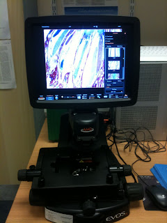
Peqlab EVOS digital fluorescent microscope

A microscope without eye pieces? Crazy?!! Not really, actually this one is pretty neat I think. We had the EVOS fl in for half a day on a demo and its really good. Nice clear images of fluorescently labelled cells acquired in a pretty short time. You can do at least 3 colours and have a phase contrast image. It uses essentially Olympus lenses that range from 2x-40x with no oil. And there is a 100x/ oil immersion available too.
It struck me as being a really good system for checking your GFP transfections, or screening your IF slides before maybe going onto taking images on more sophisticated systems. As there are no eye pieces your images are displayed on a 12" TFT screen for the world to see! Makes a good teaching tool I think, easy to explain things to students without trying to get them to look down the microscope! I guess it could be tricky to find the elusive transfected cell on a coverslip that maybe doesn't contain a great deal of them - but then that's always tricky!
I'll try and get a few images from people as examples, but for now a few iPhone snap shots:
EVOS fl, multicolour fluorescence with no eyepieces!!
The brightfield version with a colour camera

A range of lenses